Wax Models
The following are re-creations of historic teaching models, courtesy of the Mayo Clinic Historical Unit.
A history:
The models presented here are ones that have been used in classrooms, medical meetings and were on display at the Mayo Medical Museum until its closing in 1983. The models are based off of real surgeries and real patients. If necessary doctors could match the model’s number with the patient’s record and evaluate pre- and post-operative conditions.
Many of the models were also part of surgical series. These series would allow physicians and surgeons to discuss and illustrate a number of processes taking place during one surgical procedure during lectures.
Today models are still made, but are born digital as opposed to born in wax. None the less these models continue to be used in the medical school classrooms and are rotated in displays at the historical museum on Mayo Clinic’s Rochester MN campus.
Kristy Van Hoven, Mayo Clinic Historical Unit
Click on the name or image of the model to view.
Some of these models are quite graphic!
Some of these models are quite graphic!
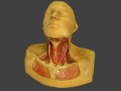
Neck Muscles

This model is used to show the various structures of neck muscles from the front.
Originally this is part of a series of surgical models used to illustrate the removal of the goiter from the patient’s thyroid tissue.
Originally this is part of a series of surgical models used to illustrate the removal of the goiter from the patient’s thyroid tissue.
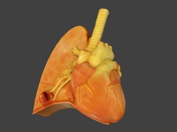
Heart and Lungs

This model illustrates the structure of the Heart and a lung including a section of the brachial tube. The heart is to the right side of the model and the lung to the left. On the lower corner of the lung there is an incision into a small growth on the lung.
This was used to illustrate the internal structure of the lung and the growth in-situ to the body.
This was used to illustrate the internal structure of the lung and the growth in-situ to the body.
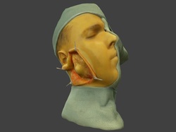
Cheek Tumor

This model represents one in a series detailing the removal of a tumor from the mandibular region. The series begins with a pre-op model of the patient, with surgical cloth around the rest of the head. Later models illustrate the various incisions, retracting locations (the small fork like structures you see are call retractors), tumor removal, and the various steps of the suture process.
This series is one of the most complex due to the nature of the procedure, but also due to the artistic detail that is required in representing the inner and outer structures of the face. You may notice this detail in the patients hair, eye brows and even eye lashes {on the original...}.
This series is one of the most complex due to the nature of the procedure, but also due to the artistic detail that is required in representing the inner and outer structures of the face. You may notice this detail in the patients hair, eye brows and even eye lashes {on the original...}.
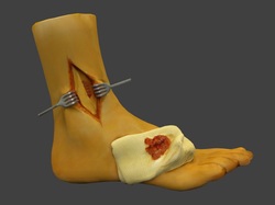
Leg Surgery

This model like many others is part of a surgical series. This model illustrates the depth of the models as well as the multi-media format. You will notice in this and other models that the creators not only used wax to create the structure of the body, but they used real surgical supplies where appropriate.
These include the gauze illustrated in this model, the surgical cloth used in the cheek model and suture thread, needles, glass syringes and other surgical instruments are used throughout the model collections.
It is these in situ details that help museum catalogers understand not only the processes discussed in historic texts, but also see how instruments were used an can detail those processes in catalog records.
For more information on the wax model collection, head over to this in-depth blog-post,
replete with many, far more graphic images.
replete with many, far more graphic images.
A big thanks to Sketchfab for their superb 3d model hosting!
If you are unable to view the models:
- Easiest solution: install and use the latest Firefox or Google Chrome. (Chrome seems to work best.)
- Refresh/reload the page (the technology is still a little fussy, sometimes).
- You don't have a compatible graphics card. Integrated graphics cards (like Intel HD Graphics) will not work well.
- You are using Internet Explorer. This is probably a deal breaker, though it may work in the future.
